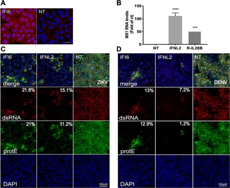
FIG 4.
Immunofluorescence of IFI6- and IFN-λ2-expressing ZIKV-infected cells. (A) Huh7 cell lines expressing IFI6 or nontargeting sgRNAs were stained with an IFI6-specific antibody Scale bar = 20 μm. (B) Medium from Huh7 cell lines expressing IFN-λ2 or NT sgRNAs was used to treat Huh7 cells. After 18 h, the cells were harvested and analyzed by qPCR using Mx1 primers. Recombinant IL-28b (IFN-λ3, R-IL-28B) was used as a positive control (10 ng/μl). ***, P < 0.01; ****, P < 0.001, one-way ANOVA Dunnett’s multiple-comparison test. (C and D) Huh7 cell lines expressing IFI6, IFN-λ2, or nontargeting sgRNAs were infected with ZIKV (MOI, 10) (C) or DENV (MOI, 1) (D) for 2 h. After 24 h (ZIKV) or 48 h (DENV), the cells were fixed and stained with flavivirus protein E-specific antibody and an antibody recognizing dsRNA. DAPI was used for nucleus staining. Scale bar = 100 μm. The relative mean fluorescence intensity of each image compared to the control was quantified using Fiji and is presented below each image.

