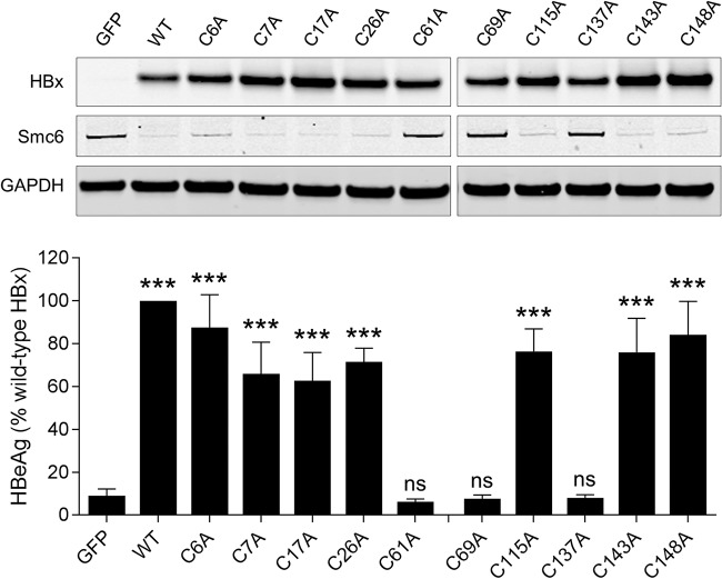
FIG 5.
Identification of cysteine and histidine residues required for HBx function in HBV-infected PHH. (Top) PHH were transduced with lentiviruses expressing GFP, wild-type HBx (WT), or HBx mutants in which a single cysteine residue was mutated to alanine. Cell lysates were analyzed by Western blotting at 72 h posttransduction. HBx was detected using an anti-Myc antibody. (Bottom) PHH were transduced with the same lentiviruses and 1 day later were infected with HBVΔX. Extracellular HBeAg was measured at day 13 postinfection. Data are expressed as a percentage of the value for the wild-type HBx control; the bar height indicates the mean of data from 3 independent experiments, and the error bars represent the SEM. Statistical significance relative to the GFP control was calculated by one-way ANOVA with Dunnett’s multiple-comparison correction. ***, P < 0.001; ns, not significant (P > 0.05).

