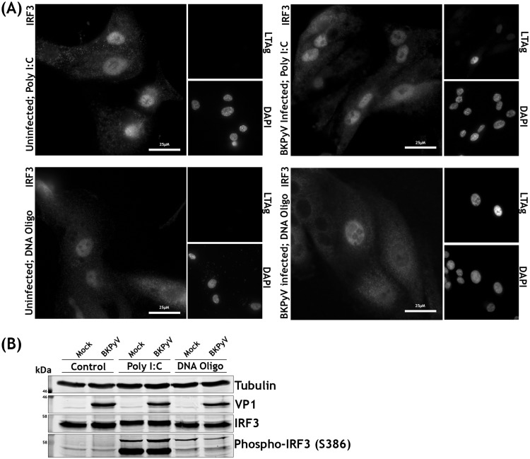
FIG 5.
BKPyV and mock-infected RPTE cells do not differ in their responses to cytoplasmic RNA and DNA. (A) Immunofluorescence microscopy analysis of IRF3 localization changes upon stimulation in BKPyV-infected or uninfected cells. RPTE cells infected with BKPyV (MOI of 0.5) or mock infected were stimulated with poly(I·C) (2 μg/ml) or oligomeric DNA (2 μg/ml) at 42 hpi and fixed at 48 hpi. DAPI was used as a nuclear marker, and anti-LTAg was used as a marker of infection. (B) Analysis of IRF3 phosphorylation upon stimulation in BKPyV-infected or uninfected cells by Western blotting. RPTE cells infected with BKPyV (MOI of 3) or mock infected were stimulated with poly(I·C) (2 μg/ml) or oligomeric DNA (2 μg/ml) at 42 hpi and harvested at 48 hpi.

