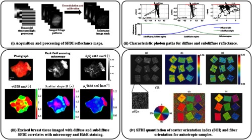
Fig. 2.
Acquisition, modeling, and application: (i) demonstration of structured illumination patterns, collection, and demodulated reflectance images—adapted from Ref. 45. (ii) Average photon paths get shorter and less diffuse with decreased source–detector separation, similar to increasing spatial frequency—adapted from Ref. 47. (iii) Subdiffuse SFDI demonstrates its ability to measure scattering-related parameters that correlate with histology of excised cancerous breast tissue—adapted from Ref. 45. (iv) SFDI demonstrates sensitivity to the amount of anisotropy (top row) and fiber orientation (bottom row)—adapted from Ref. 48.

