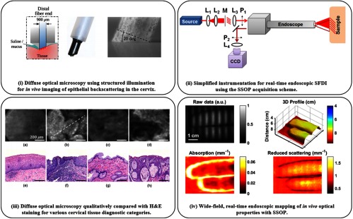
Fig. 3.
Endoscopic structured illumination imaging: (i) fiber-based probe with structured illumination for diffuse optical microscopy (DOM). (ii) 3D-SSOP enables real-time, quantitative endoscopic wide-field imaging with simplified components: lenses L, mask M, and polarizers P. (iii) (a–d) In vivo imaging using DOM of cervical tissue (e–h) with corresponding H&E histology– columns (a) and (c) are benign and columns (b) and (d) are precancerous. (iv) Endoscopic SSOP producing 3-D profile, absorption, and reduced scattering maps from a single raw frame, enabling video-rate imaging in vivo. (i) and (iii) are adapted from Ref. 79 and (ii) and (iv) are adapted from Ref. 81.

