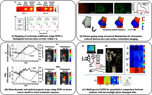
Fig. 4.
Toward clinical applications using SFDI: (i) SFDI predicts burn severity in a porcine model within 1 h using scattering coefficient imaging—adapted from Ref. 89. (ii) Photon-gating with increased spatial frequency enables absorption-reduced fluorescence imaging (top) and surface fiber orientation imaging (bottom)—adapted from Refs. 101, 102. (iii) Preclinical longitudinal study of tumor growth and chemotherapy response demonstrate feasibility for quantitative tracking of cancer therapies with SFDI—adapted from Ref. 76. (iv) Actinic skin damage assessment using mesoscopic SFDI (top-left) for mapping chromophores (top-right) and the reduced scattering coefficient (bottom) to highlight photodamage in patient P3—adapted from Ref. 116.

