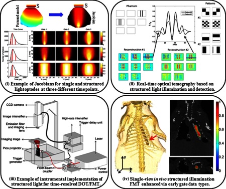
Fig. 5.
Light tomographic implementations: (i) simulated detector readings for the central source-pattern and associated Jacobians for the three gates selected: point source–point detector (top row), patterned illumination–point detector (middle row), and patterned illumination–patterned detection (bottom row adapted from Ref. 145). (ii) Simulated phantom reconstructions based on two types of patterns: phantom used for the simulations (top left), two types of patterns for illumination and detection (top right), reconstructions for each pattern type (bottom), cross sections and quantitative values of the differential absorption across a -slice. The volume for the reconstruction is divided in -slices, each having 1.25-mm thickness; even -slices are shown (top middle adapted from Ref. 146). (iii) Wide-field fluorescence lifetime imaging system for ex vivo and in vivo imaging. The schematics of the time-domain fluorescence lifetime imaging system based on a gated ICCD detection are shown (from Ref. 147). (iv) 3-D volume from the CT scan showing the position of the tube in the chest cavity (left). Coronal slice of the reconstructed volume at . (Bottom right) Transverse slice of the volume at (top right adapted from Ref. 148).

