. 2019 Jul 31;9(3):e28. doi: 10.5415/apallergy.2019.9.e28

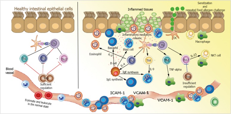
Fig. 3. Abnormal interactions between allergen and the host immune system in gastrointestinal tissue in eosinophilic esophagitis (EoE) pathogenesis. Antigen presenting cells present food allergens as major histocompatibility complex class II conjugates to T-cell receptors on naive T cells. T cells then differentiate into T-helper type 1 (Th1) or Th17 cells, which secrete different pro-inflammatory and anti-inflammatory cytokines leading to recruitment of humoral and cellular factors of innate immunity. In healthy tissues (left), immune homeostasis is maintained by interdependent control exerted by sufficient Treg cells. In mucosal response in EoE (right), dysfunctional regulation of Th1 or Th17 pathways triggers an unregulated inflammatory response and recruitment of innate immune cells. Cytokine levels elevation can promote oxidative tissue damage and facilitate proteolytic peptides and enzymes recruitments, eventually presenting as gastrointestinal disease. Furthermore, enactment Th2 pathway can prompt plasma cells identifying fungal cell wall antigens and delivering antiglycan antibodies production. IL, interleukin; NKT, natural killer T; ICAM, intercellular adhesion molecule; VCAM, vascular cell adhesion molecule.

Fig. 3