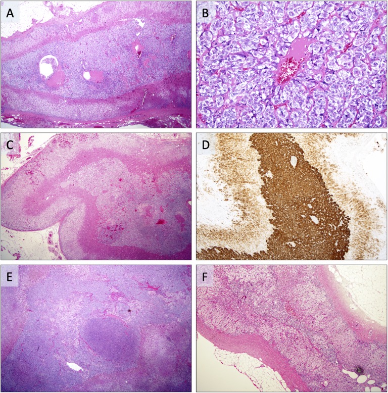
Figure 2.
Photomicrographs of adrenal specimen with AMH. (A) AMH represented by a substantial widening of the adrenal medulla in a patient with synchronous PCC. (B) Same case magnified ×200, demonstrating chromaffin cells without pleomorphism arranged in small lobules. (C) Patient with MEN2A and a germline RET mutation, displaying AMH with concurrent PCC. Note the augmented widening of the medulla. (D) Same case positive for synaptophysin, with strong cytoplasmic immunoreactivity in the AMH and weaker expression in the adjacent adrenal cortex. (E) Patient with synchronous PCC displaying AMH with distinct micronodular arrangements. (F) Unrelated reference case without AMH. Note the width of the adrenal medulla compared with photomicrographs A–E.

