Figure 1.

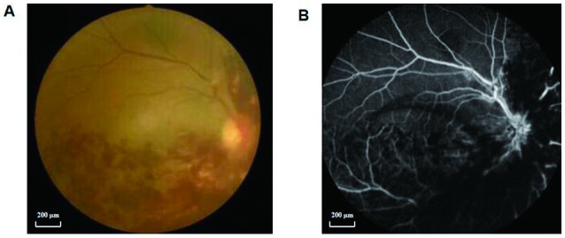
Example of RVO seen on fundus colorized photography and FFA. (A) Colorized image of the fundus of an eye with RVO. (B) FFA of the same eye with RVO. RVO, retinal vein occlusion; FFA, fundus fluorescein angiography.

Example of RVO seen on fundus colorized photography and FFA. (A) Colorized image of the fundus of an eye with RVO. (B) FFA of the same eye with RVO. RVO, retinal vein occlusion; FFA, fundus fluorescein angiography.