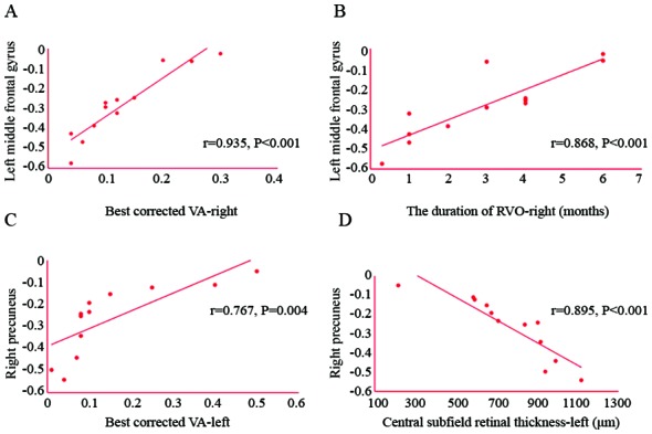
Figure 5.

Correlation between the mean ALFF values of different brain areas and clinical behavior. (A) The best-corrected VA in the left eye was positively correlated with the ALFF value of the right precuneus (r=0.767, P=0.004). (B) The duration of RVO in the right eye was positively correlated with the ALFF value of the left middle frontal gyrus (r=0.868, P<0.001). (C) The best-corrected VA in the right eye was positively correlated with the ALFF value of the left middle frontal gyrus (r=0.935, P<0.001). (D) The central subfield retinal thickness in the left eye was negatively correlated with the ALFF value of the right precuneus (r=−0.895, P<0.001). RVO, retinal vein occlusion; VA, visual acuity. ALFF, amplitude of low-frequency fluctuation.
