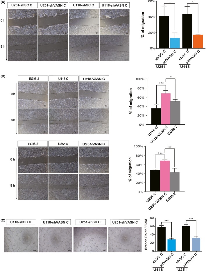
Figure 6.

Vasorin (VASN)‐expressing tumor cells strongly promoted HUVEC migration and tube formation. Micrographs and histograms of the scratch wound‐healing assay of HUVEC grown in conditioned medium (CM) from glioma cell knockdown (A) and overexpression (B) of VASN. EGM‐2 served as a control. C, Micrographs of tube formation by HUVEC grown in CM from shVASN‐transfected glioma cells as indicated. Quantification of the number of branch points formed during the tube formation assay. Mean number of branching points per ×100 field ±SEM. *P < 0.05, **P < 0.01, ***P < 0.001. Scale bar, 100 μm
