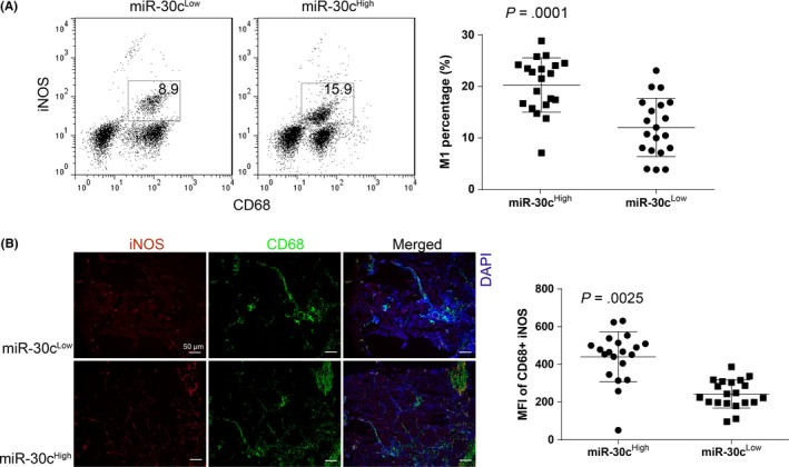
Figure 3.

High expression of microRNA (miR)‐30c is related to the increased percentage of M1 cells within tumor‐associated macrophages (TAMs). A, Percentage of M1 cells (defined as CD68+ and inducible nitric oxide synthase [iNOS]+) with gastric cancer (GC) TAMs were determined by using flow cytometry. The comparison was undertaken between miR‐30cLow and miR‐30cHigh GC TAMs. B, Percentage of M1 cells with GC TAMs was determined by using immunofluorescence staining (×100), the integrated optical density value of each figure was obtained by using Image J analyzing 5 random visions. All experiments were undertaken in triplicate. Data are presented as mean ± SD. Student's t test. MFI, mean fluorescence intensity
