Figure 2.

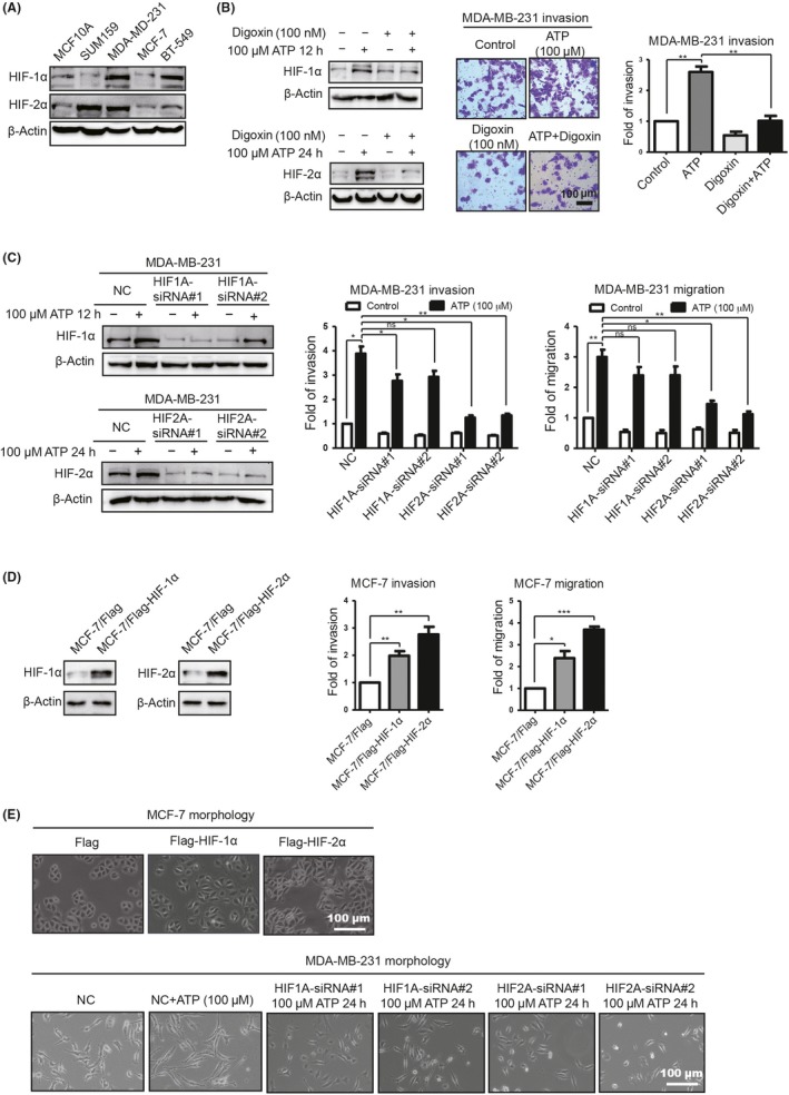
Extracellular ATP promotes breast cancer cell invasion via hypoxia‐inducible factor (HIF)‐2α in vitro. A, HIF‐1/2α expression was examined by western blotting. B, Western blotting demonstrated that HIF‐1/2α was decreased by digoxin (left). Transwell invasion assays illustrated that ATP‐driven invasion was attenuated (right). C, Western blotting demonstrated that HIF‐1/2α was knocked down (left). Transwell invasion and migration assays showed that ATP‐driven invasion and migration were attenuated via HIF2A‐siRNA (right). D, Western blotting illustrated HIF‐1/2α elevation in stably transfected MCF‐7 cells (left). Transwell invasion and migration assays showed that HIF‐1/2α enhanced MCF‐7 invasion and migration capacities. E, The morphology of MCF‐7/Flag‐HIF‐2α cells looked like dispersed spindle‐shaped mesenchymal cells, while MCF‐7/Flag cells displayed cobblestone‐like clusters of epithelial cells (upper). The shape of the MB‐231/HIF2A‐siRNA‐treated cells looked more like the shape of epithelial cells (lower). Error bars represent means ± SD from triplicate experiments. *P < 0.05; **P < 0.01; ***P < 0.001; ns, not significant
