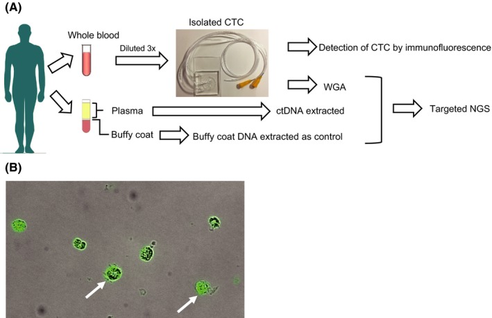
Figure 1.

Flow diagram of the optimized protocol for detecting genomic alterations in circulating tumor cells (CTC) and ctDNA, and immunofluorescence cytochemistry of isolated CTC. A, Blood from patients (up to 2 × 5 mL of peripheral blood) was collected using EDTA vacutainers. One collection tube of hemolyzed whole blood was diluted 3‐fold. CTC were isolated from the blood using a label‐free inertial microfluidics approach (LFIMA). ctDNA and buffy coat DNA were isolated from the other collection tube. Targeted next‐generation sequencing was performed using the extracted DNA. B, Fluorescence image of isolated CTC stained for cytokeratin (green). NGS, next‐generation sequencing; WGA, whole‐genome amplification
