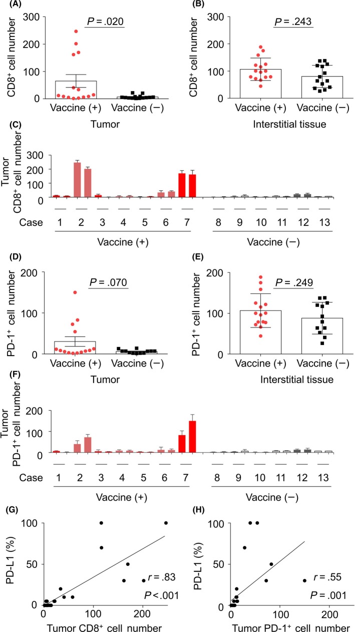
Figure 3.

In situ investigation of infiltration of CD8+ and programmed cell death‐1 (PD‐1)+ lymphocytes into pancreatic carcinoma lesions. A,B, Comparison of infiltration of CD8+ cells in the tumor (A) and interstitial tissue (B) between patients who had and had not received the survivin 2B peptide (SVN‐2B) vaccine. C, Number of CD8+ lymphocytes that infiltrated each tumor lesion. D,E, Comparison of infiltration of PD‐1+ cells into the tumor (D) and interstitial tissue (E) between patients who had and had not received the SVN‐2B vaccine. F, Number of PD‐1+ lymphocytes that infiltrated each lesion. G,H, Correlation between proportions of the PD‐L1+ tumor and CD8+ (G) or PD‐1+ (H) lymphocyte infiltration in the lesions. PD‐L1 expression on the surface of tumor cells was deemed to be positive. Signal intensity was not evaluated
