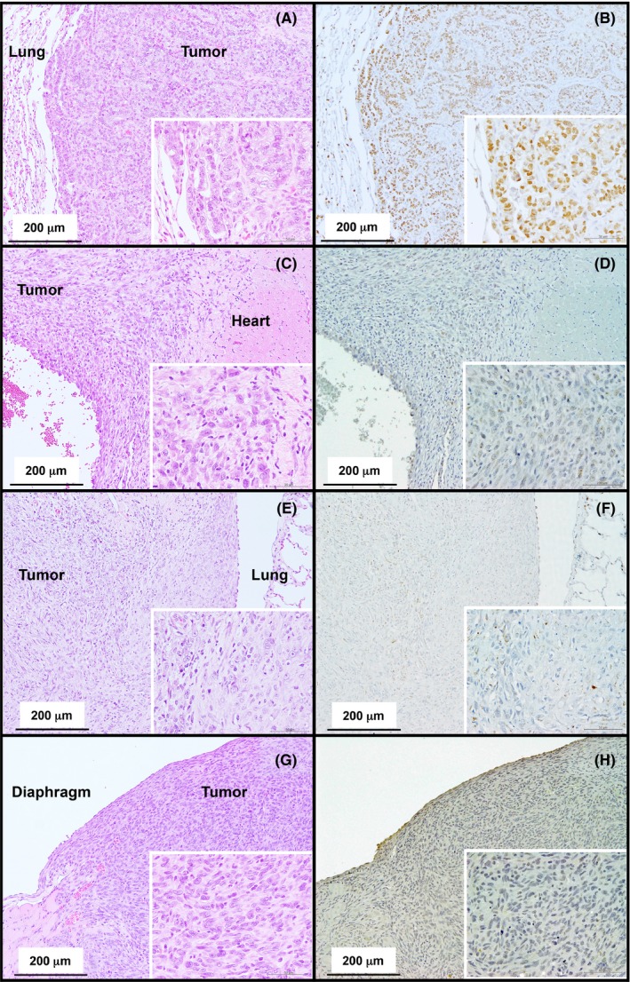
Figure 3.

Representative histopathological images of the lung adenocarcinoma in the vehicle‐treated rat (A) and mesotheliomas that developed in the MWCNT‐7 treated rats (C, E, and G). Immunohistochemical staining of TTF‐1 (B, D, F and H). Nuclei in the adenocarcinoma are positive for TTF‐1 (B). The malignant mesotheliomas near the heart, thoracic cavity, and diaphragm are negative for TTF‐1 (D, F, and H)
