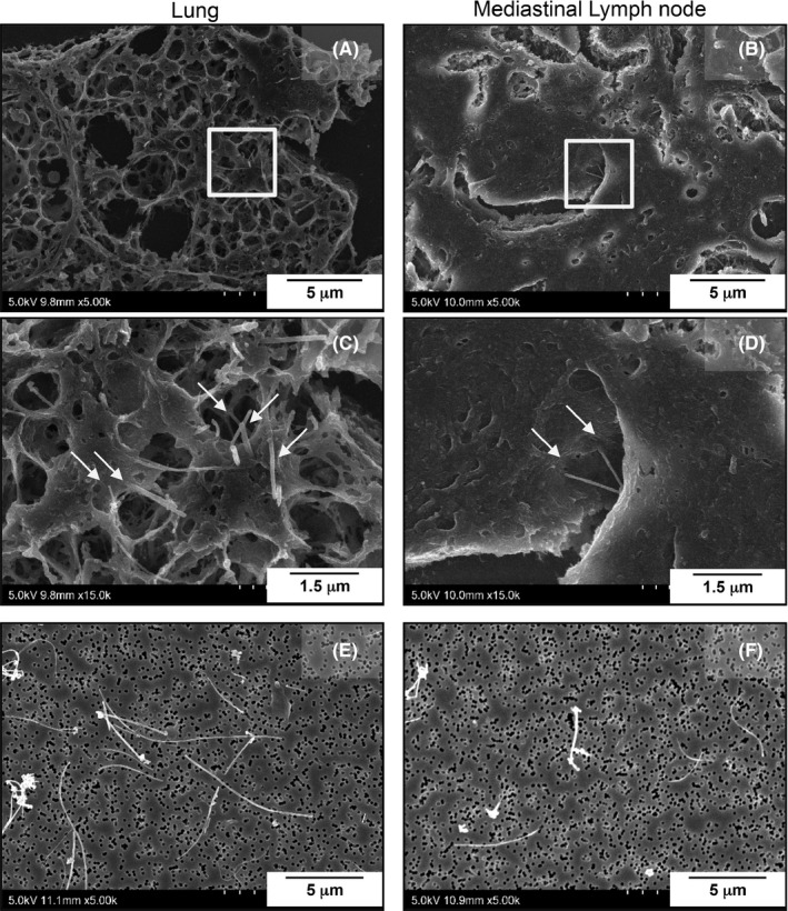
Figure 4.

A‐D, Scanning electron microscopic images of MWCNT‐7 fibers (arrows) phagocytosed by macrophages in the lung and mediastinal lymph node. E‐F, representative images of MWCNT‐7 fibers from the lung and mediastinal lymph node after tissue digestion
