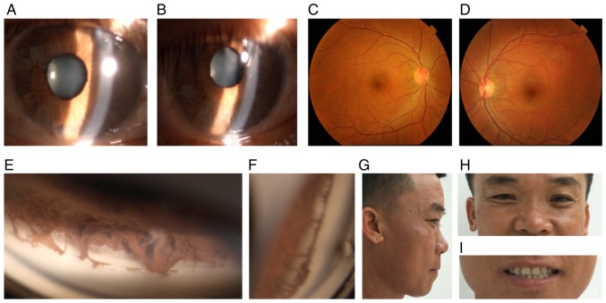
Figure 3.
Ocular characteristics and systemic anomalies of patient II:2. (A) Slit lamp images of the right and (B) left eye revealed atrophy of the iris. (C) The fundus of the right and (D) left eye were normal. (E) Gonioscopy indicated open angles in the right and (F) left eye, with prominent iris processes and anterior insertion of the iris into the trabecular meshwork. (G and H) Physical examination revealed midface hypoplasia, hypertelorism, telecanthus and flat broad nasal bridge. (I) The dentition was normal.

