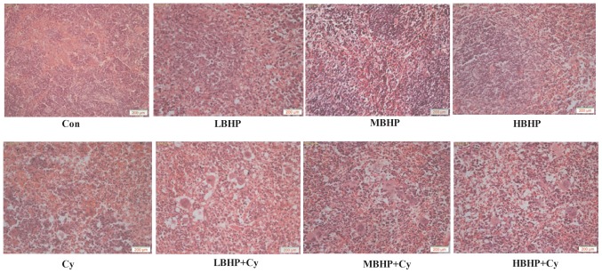
Figure 5.
Hematoxylin-eosin staining of the mouse spleen. Images were captured at ×100 magnification with a light microscope. The spleen of the mice in the Cy group, LBHP + Cy group, MBHP + Cy group and HBHP + Cy group all exhibited abnormalities to varying degrees compared with that in the Con group. The lymphocyte arrangement was sparse, and the structure was ambiguous and irregularly arranged. The number of lymphocytes was decreased, red veins were engorged with small veins, the splenic sinus was marginally expanded, and lymphoid follicles and necrotic cells were visible. The Cy group exhibited the most marked changes. No substantial changes were found among the LBHP group, MBHP group and HBHP group. The LBHP + Cy group, MBHP + Cy group and HBHP + Cy group had relatively neatly arranged lymphocytes and relatively clear cell structure. All data were from two independent experiments, each of which was repeated in parallel twice. BHP, bioactive hepatic peptide; LBHP, low-dose BHP; MBHP, mid-dose BHP; HBHP, high-dose BHP; Cy, cyclophosphamide.

