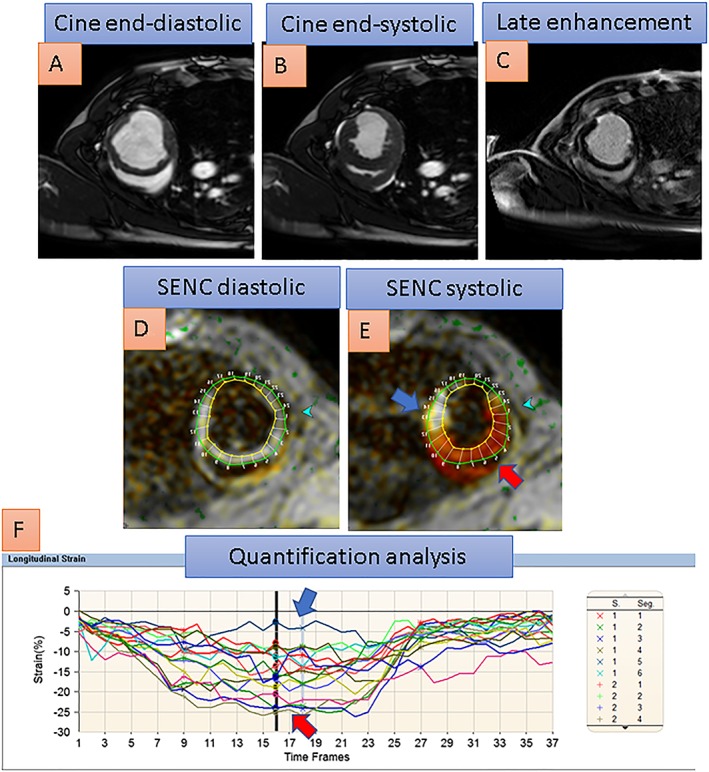
Figure 2.

The midventricular short axis view of a porcine heart with akinesia of the lateral left ventricular wall after infarction due to closure of its circumflex coronary artery can be appreciated with cine images in A and B. With conventional SENC images, red colour corresponds to normal contracting myocardium in the septal wall, faded orange and yellowish colour corresponds to reduced strain in peri‐infarct areas, and white colour corresponds to severely reduced or absent myocardial contraction in the infarcted lateral wall. Late gadolinium enhancement (in C) exhibits 75% transmural infarction of the lateral wall, which corresponds to severely reduced strain, coded white in the lateral wall (blue arrow) compared with normal strain, coded red in the septal wall (red arrow), with the systolic SENC image in E. Quantification analysis in F reveals severely reduced systolic strain in the lateral wall segments (blue arrow in F) compared with normal strain in septal wall segments (red arrow in F).
