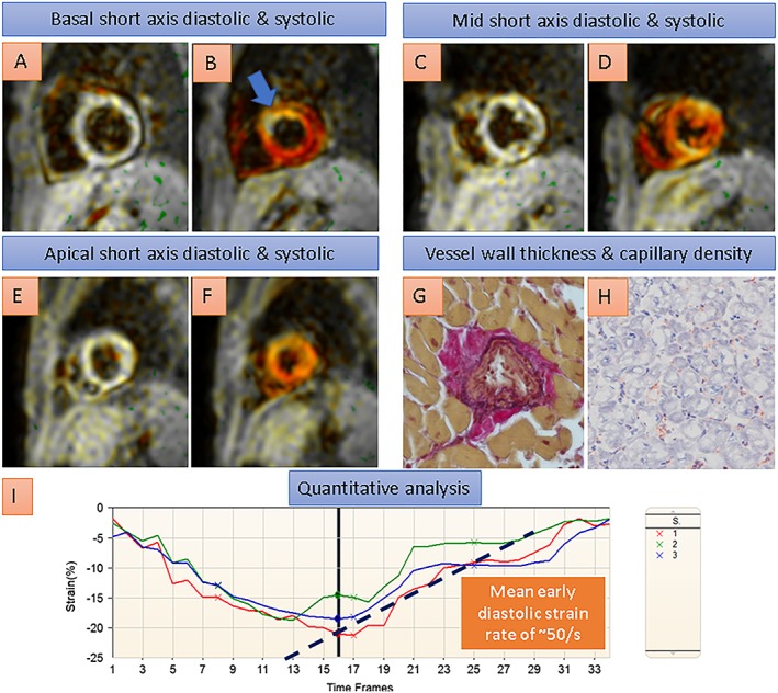
Figure 5.

A heart transplant recipient with normal coronary arteries by angiography and normal ejection fraction of 55% by cardiovascular magnetic resonance. In this patient, conventional SENC acquisitions depicted reduced systolic strain in the basal anterior wall (blue arrow in B, coded yellowish/white compared with normal strain, e.g. in the inferior wall, coded red), and reduced diastolic strain rates, indicating impaired diastolic left ventricular function, were demonstrated by quantitative analysis of the strain curves in I. Endomyocardial biopsy showed thickened coronary arterioles and reduced capillary density (G and H) by histologic criteria, consistent with transplant microvasculopathy.
