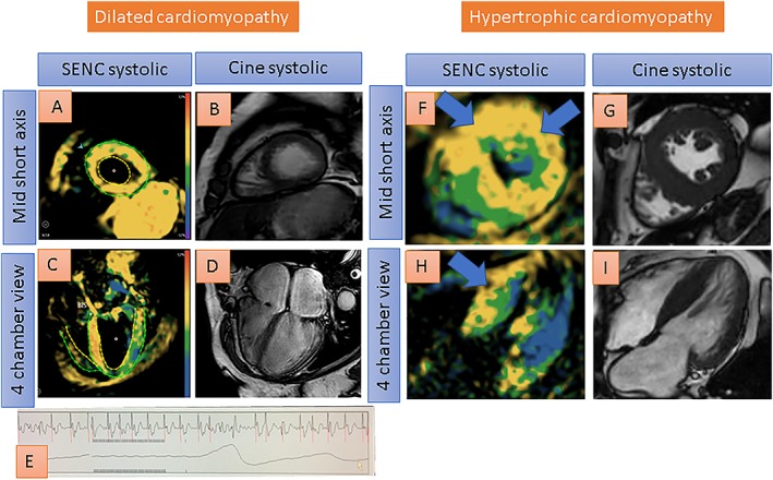
Figure 6.

Single‐heartbeat fast‐SENC acquisitions of a patient with dilated cardiomyopathy (A–E) and another patient with hypertrophic cardiomyopathy (F–I). Despite atrial fibrillation, good image quality is provided, depicting globally reduced strain using fast‐SENC in A and C (reduced strain coded green or yellow by fast‐SENC acquisitions). Ejection fraction is ~20% with cine images in B and D. The electrocardiogram shows atrial fibrillation (E). Severe left ventricular hypertrophy, on the other hand, can be appreciated with fast‐SENC (F and H) and with systolic cine images (G and I) in another patient with hypertrophic cardiomyopathy. Reduced strain especially in the septum and in the anterior wall (coded yellow) can be depicted with fast‐SENC acquisitions (blue arrows in F and H).
