
Figure 8.
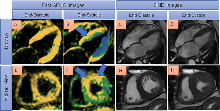
Reduced myocardial strain can be depicted by single‐heartbeat fast‐SENC acquisitions especially in the anterior and septal wall (blue arrows in B and F, severely reduced myocardial strain coded yellow) in a patient with hypertensive and diabetic heart disease. Four chamber view and short axis cine images (C and D, and G and H) show preserved ejection fraction of 58% in the presence of evident left ventricular hypertrophy.