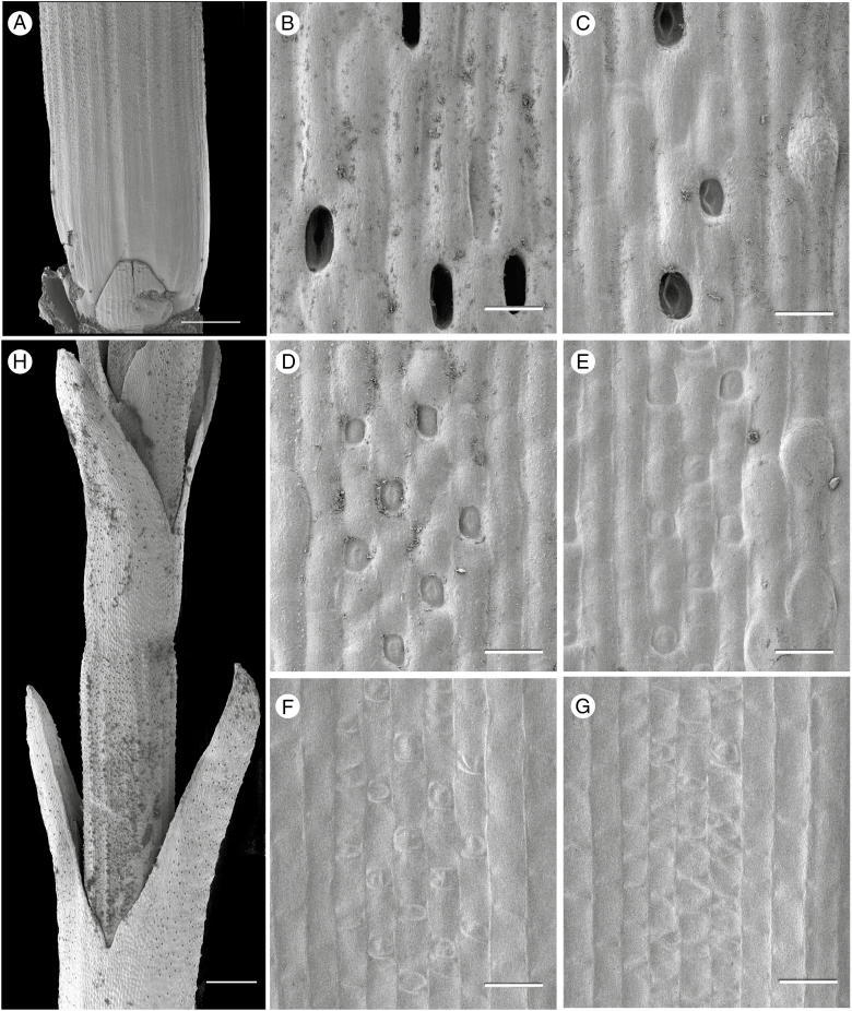
Fig. 2.
Ephedra likiangensis (SEM). (A) Stem internode with scale leaves removed from node, revealing an axillary bud (at base). (B–G) Details of surface of stem shown in (A) from top to bottom, showing series of stomatal developmental stages across a single internode, from mature sunken stomata in (B) to meristemoids in (G). (H) Internode with congenitally fused pairs of scale leaves at each node. Scale bars: (A, H) = 500 μm; (B–G) = 20 μm.

