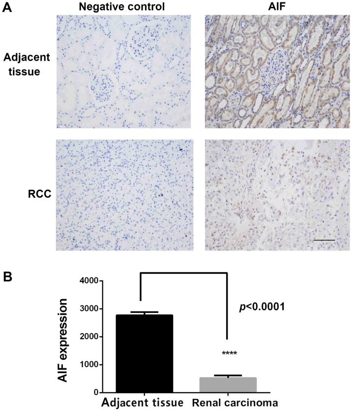
Figure 1.
AIF expression in RCC and adjacent normal tissues. (A) IHC staining of AIF expression in RCC and adjacent tissues. Strong staining of AIF was observed in normal kidney sections compared with weak staining in the adjacent tissues. (B) Quantification of AIF staining in IHC images of RCC and adjacent normal tissues. AIF expression was significantly decreased in RCC tissues. ****P<0.0001, n=96. Scale bar, 200 µm. AIF, apoptosis-inducing factor; RCC, renal cell carcinoma; IHC, immunohistochemistry.

