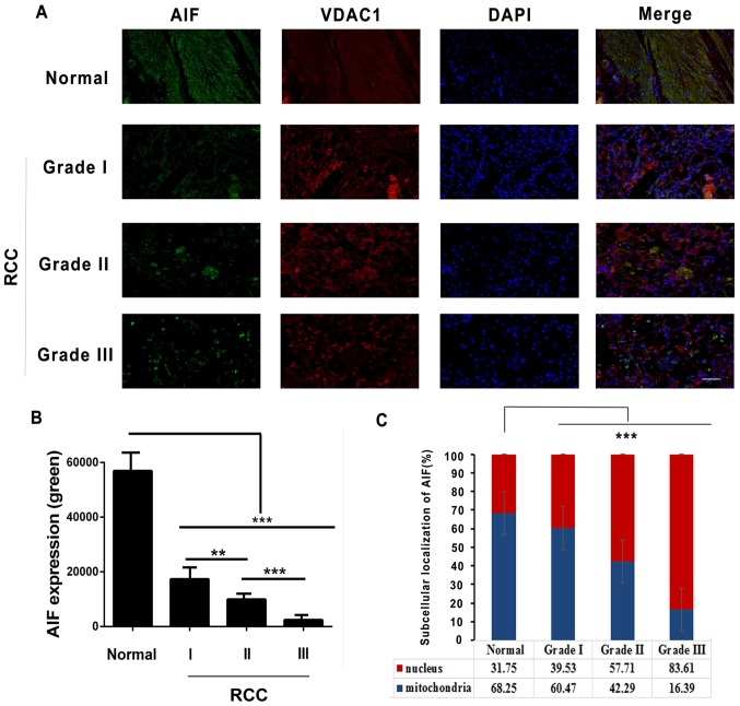
Figure 4.
Subcellular localization of AIF in RCC cells. (A) Immunofluorescence staining of RCC grades I, II and III and adjacent normal tissue with antibodies against AIF (green), VDAC1 (red; mitochondrial marker) and with DAPI (blue; nuclear stain). (B) Quantification of AIF expression in RCC and adjacent normal tissue. (C) Quantification of AIF staining in mitochondrial and nuclear subcellular compartments in RCC grades I, II and III and adjacent normal tissue. **P<0.01, ***P<0.001 (n=6). Scale bar, 500 µm. AIF, apoptosis-inducing factor; RCC, renal cell carcinoma; VDAC1, voltage-dependent anon-selective channel 1.

