Fig. 1.

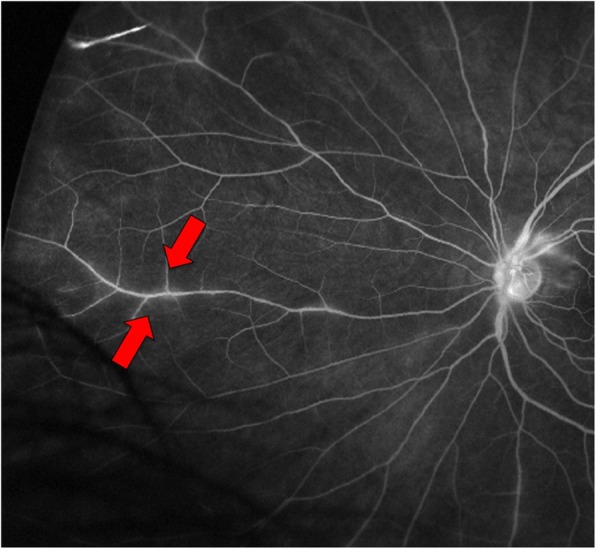
Fluorescein angiogram of the left eye demonstrating regions of retinal vascular staining and leakage near the optic nerve and in the peripheral retina (arrows)

Fluorescein angiogram of the left eye demonstrating regions of retinal vascular staining and leakage near the optic nerve and in the peripheral retina (arrows)