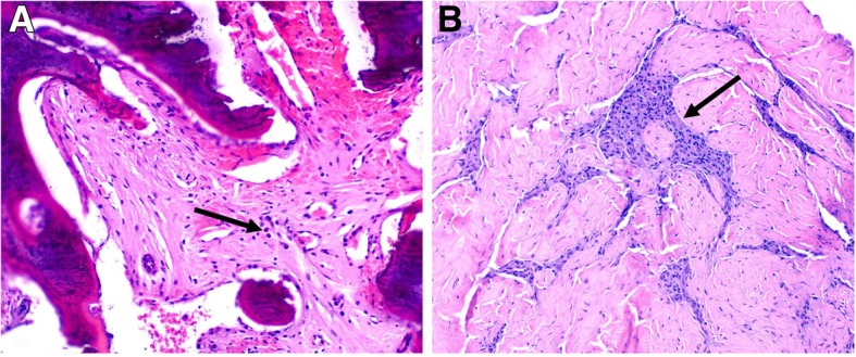
Fig. 3.

a Sternum biopsy showing thickened bony trabeculae (incompletely decalcified during tissue processing) and fibrosis in the medullary cavity with small collections of plasma cells (arrow) and scattered lymphocytes (H&E stain, X100). b Sternoclavicular joint biopsy showing dense fibrous connective tissue, presumed to be the joint capsule, with focal collections of lymphocytes and plasma cells (arrow). (H&E stain, X100)
