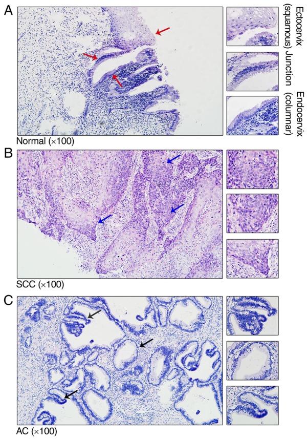
Figure 1.
Representative histological haematoxylin and eosin staining of the adult cervix. (A) Normal cervical tissue with squamous, ectoendocervical squamocolumnar junction and columnar cells (red arrows). (B) SCC cervical tissue with representative nidus (blue arrows). (C) AC cervical tissue with representative nidus (black arrows). The magnified views (×200) are representative structures, which are indicated by the arrows. SCC, squamous cell carcinoma; AC, cervical adenocarcinoma.

