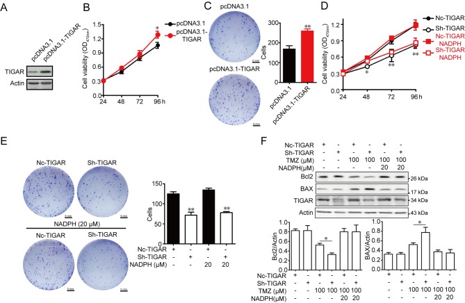
Figure 3.
Effect of TIGAR on viability and apoptosis in U-87MG glioma cells. (A) TIGAR content was increased after transfection. (B) Overexpression of TIGAR promoted viability in U-87MG cells, as measured by am MTT assay (n=5). (C) Cells (200) transfected with pcDNA3.1 and pcDNA3.1-TIGAR were seeded into 6-well plates and cultured for 14 days. Colony formation was detected using crystal violet staining. The colony number was counted (n=3). Scale bar, 5 mm. (D) NADPH treatment of TIGAR-knockdown U-87MG cells had no effect on viability (n=5). (E) NADPH treatment of TIGAR-knockdown U-87MG cells had no effect on colony formation (n=3). Scale bar, 5 mm. (F) NADPH (20 µM) addition decreased the expression levels of BAX protein and increased Bcl2 protein expression in 100 µM TMZ-treated TIGAR knockdown U-87MG cells. β-actin was used as an internal control. Bar graphs depict semi-quantitative analysis of Bcl2 and BAX expression (n=3). Data are expressed as the means ± standard deviation. *P<0.05, **P<0.01 vs. pcDNA3.1 or NC-TIGAR. BAX, Bcl2-associated X protein; Bcl2, B cell lymphoma 2; NADPH, reduced nicotinamide adenine dinucleotide phosphate; NC, negative control shRNA; OD, optical density; sh, short hairpin RNA; TIGAR, TP53 induced glycolysis regulatory phosphatase; TMZ, temozolomide.

