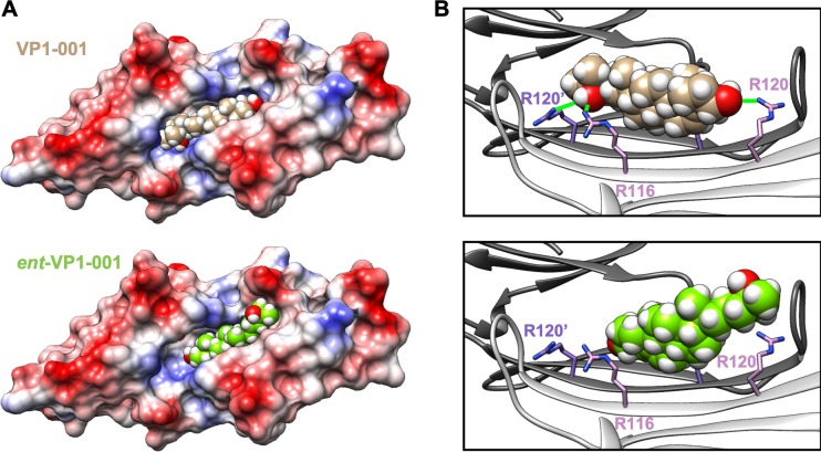
Figure 2.
VP1-001 binds to the ACD, but ent-VP1-001 does not. (A) Electrostatic surface model of the ACD dimer in complex with docked VP1-001 (top) or ent-VP1-001 (bottom). Note the relatively poor ability of ent-VP1-001 to fit into the pocket. (B) Ribbon views of the ACD pocket with bound VP1-001 (top) or ent-VP1-001 (bottom). Green lines (top) indicate predicted hydrogen bonds (distance 4 Å or less). The protein coordinates are from the x-ray structure 2WJ7. However, the angle between protomers (shown in white and gray) was reduced by 8° by a rigid body movement (as described in Methods).

