Figure 7.
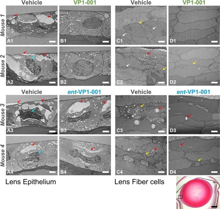
VP1-001, but not ent-VP1-001, improves lens morphology in cryAB(R120G)-mutant mice. Electron micrographs of lens epithelium and lens fiber cell sections from four mice are grouped horizontally (for example, images A1, B1, C1, and D1 are from one mouse). Scale bars: 2 μm, white. Red arrows indicate vacuoles, cyan arrows indicate an abnormal nuclear shape, yellow arrows indicate irregular cell size, and white arrows indicate abnormal cell protrusions. (Left) Lens epithelial sections from treated and untreated eyes of cryAB(R120G) heterozygous mice. For each treatment condition, two replicate mice are displayed. (Right) Lens fiber sections from the same four mice. The images are representative of a majority of the sections from mice. In contrast to VP1-001, the enantiomer did not reverse morphologic changes in cryAB(R120G) heterozygous mice. (Bottom panel) Low-magnification image of a lens treated with VP1-001. The equatorial region in which the EM images were taken is enclosed by a box. Scale bar: 0.5 mm, black.

