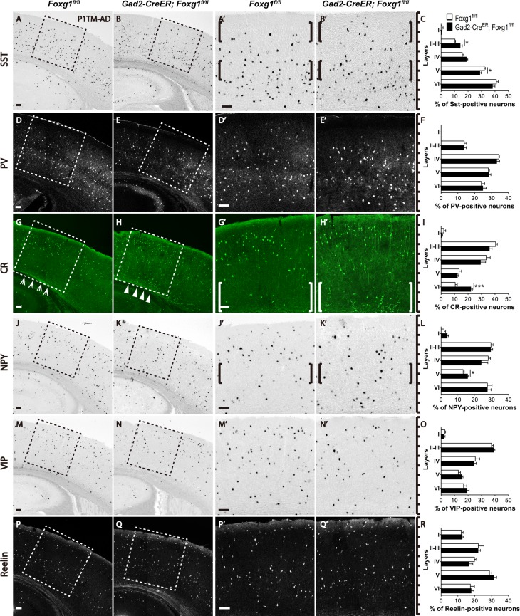
Figure 2.
The SST-, CR-, NPY-positive subtype interneurons exhibited abnormal distributions in the adult cortex after Foxg1 ablation. (A–C) SST-positive interneurons were preferentially located in the deeper layers in the controls (A, A’, C), but more SST-positive interneurons were located in upper layer II–III, with a decreased number in layer V (B, B’ C; Control, n = 5; MU, n = 4; layer II–III, P = 0.0341; layer V, P = 0.0316). (D–F) The distribution of PV-positive interneurons in the mutants (E, E’, F) was not altered compared with the controls (D, D’, F; Control, n = 5; MU, n = 4). (G-I) More CR-positive interneurons accumulated in the deeper layers in the mutants (H, H’, I) than in the controls (G, G’, I; Control, n = 5; MU, n = 4; layer VI, P = 0.0006). (J–L): More NPY-positive interneurons were distributed in layer V in the mutants (K, K’, L; Control, n = 3; MU, n = 3; layer V, P = 0.0265). (M–R) No alterations were detected in the distributions of VIP-positive interneurons (M–O; Control, n = 4; MU, n = 4) and Reelin-positive interneurons (P–R; Control, n = 4; MU, n = 4). A’, B’, D’, E’, G’, H’, J’, K’, M’, N’, P’ and Q’ show higher magnification images of the boxed areas in A, B, D, E, G, H, J, K, M, N, P and Q, respectively. Scale bar: 100 μm. Student’s t-test, *P < 0.05, **P < 0.01.

