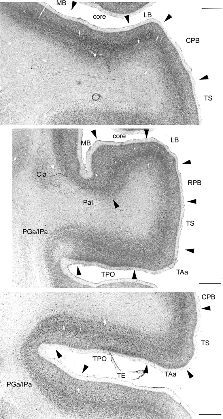
Figure 2.
Low power photomicrographs showing the cytoarchitectural criteria used to distinguish areas in the superior temporal cortex of Cebus. Cytoarchitectural transitions are indicated by arrowheads. Top: Section through the lower bank of the lateral sulcus and superior temporal gyrus, at the level of the primary auditory cortex (part of the auditory core), showing transitions to the lateral belt (LB) and MB, as well as the characteristics of the caudal parabelt (CPB) and cytoarchitectural area TS. Middle: Rostral section, at the level of the rostral auditory field (indicated as “core”). The transitions between the LB and MB areas to the rostral parabelt (RPB) and parainsular (PaI) cytoarchitectural areas are indicated.TAa, TPO, and PGa/IPa are cytoarchitectural areas defined by Pandya and colleagues in the macaque (e.g., Seltzer and Pandya 1978). Cla—claustrum. Bottom: A section at a level similar to the one shown in the top panel, showing the transitions between polysensory areas in the superior temporal gyrus and sulcus. Scale bars = 1 mm.

