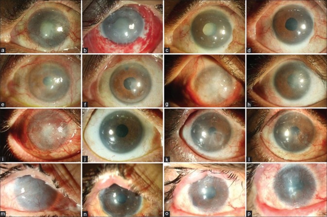
Figure 4.
Clinical outcomes of allogenic simple limbal epithelial transplantation (SLET). The top row shows the 1-year progressive outcomes in a case of mucous membrane pemphigoid (MMP) with advanced senile cataract (a to d). Preoperative image showing total limbal stem cell deficiency (LSCD) with mature senile cataract (a); postoperative day (POD) 1 image showing intact transplants on the cornea with multiple hemorrhages under the human amniotic membrane graft (b); POD 90 image showing a epithelized avascular cornea, at this visit the patient was planned for cataract surgery (c); 12 and 9 months after allogeneic SLET and cataract surgery, respectively, the aided visual acuity is 20/20 for distance and n6 for near (d). Pre- (e) and postoperative (f) 1-year images of a one-eyed patient with OSSN excision–induced LSCD. Pre- (g) and postoperative (h) 1.5-year images of a case of bilateral LSCD due to severe chronic ocular allergy. The third row from top summarizes the 2-year timeline of another case of MMP with total LSCD (i) where a successful outcome was maintained until 1.5 years, following which there was an episode of immunological rejection (k) which was reversed but the patient developed partial LSCD (l). The bottom row shows the 4-year timeline of a case of bilateral LSCD (m) due to Stevens–Johnson syndrome (SJS) who first underwent lid-margin mucous membrane grafting followed by allogeneic SLET and maintained a stable surface (n) for 2.5 years following which he gradually developed recurrence of LSCD (o-p)

