
Figure 1.
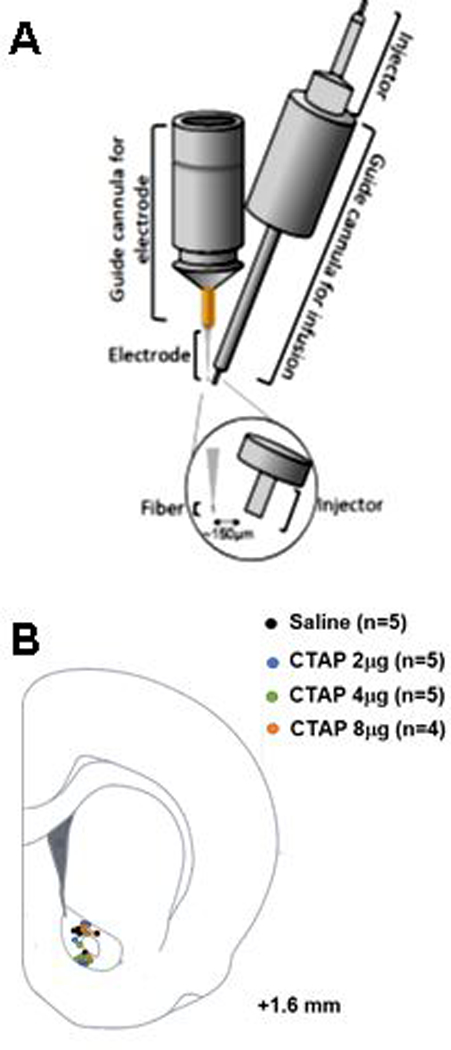
Schematic representation of the guide cannula ensemble and anatomical placements for simultaneous in vivo voltammetric measurement of evoked dopamine release and local application of a drug. (A) Guide cannula ensemble: The electrochemical measurements were performed at carbon-fiber microelectrodes lowered via the guide cannula on the left. The drug applications were performed via the injector inserted into the guide cannula on the right. Both cannulae were positioned and cemented together under a microscope prior to any experiment to ensure an approximate distance of 150 μm between the electrode and end of the injector. (B) Representations of electrode/injector placements within nucleus accumbens core.