Figure 2:
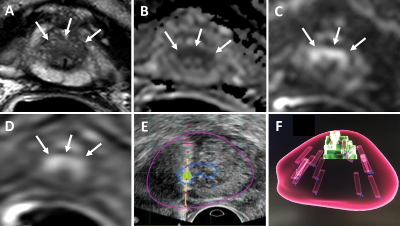
Images in a 75-year-old man with a serum prostate-specific antigen level of 14.6 ng/mL. A, T2-weighted MRI, B, apparent diffusion coefficient map, C, diffusion-weighted MRI (b value, 2000 sec/mm2), and, D, dynamic contrast-enhanced MRI show a lesion (arrows) classified as Prostate Imaging Reporting and Data System category 4 in A and C and as positive in D in the midline anterior apical transition zone. E, Targeted biopsy was performed with transrectal US/MRI and revealed Gleason grade 3+4 cancer in three of the four cores sampled from this lesion. F, Volume-rendered MRI shows mapping of the four targeted cores and systematic 12 cores. One of the 12 systematic biopsy cores revealed Gleason grade 3+3 prostate adenocarcinoma (5% core involvement), which made no difference for risk stratification in this patient.

