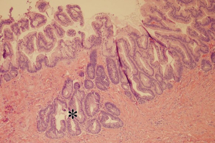
Figure 5f.
Mucinous borderline tumor in a 63-year-old woman with minimal abdominal pain and urinary frequency. (a) Sagittal US image shows a cystic mass (*) superior and anterior to the bladder (BL). (b–d) Sagittal T2-weighted (b), coronal T2-weighted (c), and coronal contrast-enhanced fat-saturated T1-weighted (d) images show a cystic mass (*) with minimal complexity (arrow in c and d). (e) Photograph of the gross pathologic specimen shows a mass with minimal complexity (black arrow) filled with thick fluid (white arrow). (f) High-power photomicrograph shows epithelial proliferation, stratification, and atypia (*), consistent with mucinous borderline tumor. (H-E stain.)

