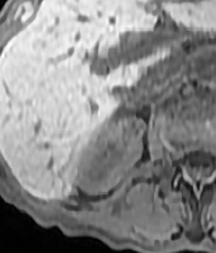
Figure 4c:

Axial images in 73-year-old woman with lung cancer and an indeterminate right renal mass identified at staging CT. (a) Unenhanced and (b) nephrographic phase CT images show a nonenhancing mass with thick calcifications. Because of the potential for thick calcifications to obscure underlying enhancing features, MRI was performed. (c) Unenhanced, (d) nephrographic, and (e) subtraction images from T1-weighted gradient-echo MRI show few (≤3) thin (≤2 mm) enhancing septa. This mass would be considered Bosniak IIF in the current classification (because of the thick calcifications) and Bosniak II in the 2019 update. During surveillance, the mass has remained unchanged for more than 11 years.
