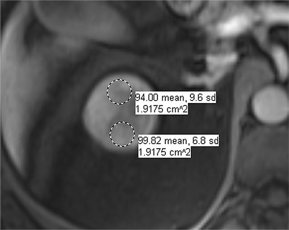
Figure 5h:

Images in 66-year-old man with prostate cancer and an indeterminate right renal mass. (a) Unenhanced and (b) axial nephrographic CT images show a nonenhancing heterogeneous mass. Because of the heterogeneity of the mass, MRI was performed. At (c) coronal T2-weighted single-shot fast spin-echo MRI, the mass was hypointense to the renal cortex. (d) Axial image from in-phase (right) dual-echo gradient-echo MRI shows signal loss relative to that on shorter-echo-time opposed-phase axial image (left), indicating the presence of hemosiderin. sd = Standard deviation At (e) axial fat-saturated unenhanced T1-weighted spoiled gradient-echo MRI, the mass was heterogeneously hyperintense. At (f) axial postcontrast MRI performed during the nephrographic phase with the same acquisition parameters as e, there was no visible enhancement. Therefore, (g, h) regions of interest were placed, and they revealed subtle nodular enhancement (a ≥ 15% signal intensity increase between the unenhanced [g] and nephrographic [h] phases). Because nodular enhancement was detected, the mass would be classified as a Bosniak IV mass. The mass was resected and was found to be a hemorrhagic papillary renal cell carcinoma. (Note that if no enhancing features had been identified, the mass would be classified as Bosniak IIF in the updated classification because of its heterogeneous hyperintensity at fat-saturated T1-weighted MRI.) sd = Standard deviation.
