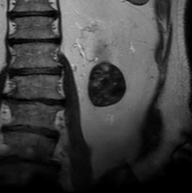
Figure 6b:
Images in 68-year-old man with an indeterminate left renal mass. (a) Axial unenhanced (left) and nephrographic phase (right) CT images show a partially calcified nonenhancing heterogeneous mass. Because of the heterogeneity of the mass, MRI was performed. At (b) coronal T2-weighted single-shot fast spin-echo MRI, the mass was hypointense to the renal cortex. At (c) coronal T1-weighted fat-saturated spoiled gradient-echo MRI, the mass was heterogeneously hyperintense. (d, e) At coronal dynamic postcontrast subtraction MRI in the (d) corticomedullary and (e) nephrographic phases, there was unequivocal visible enhancement within multiple nodules (eg, arrow in e). Because nodular enhancement was detected, the mass would be classified as Bosniak IV. The mass was biopsied and was found to be a hemorrhagic papillary renal cell carcinoma. (Note that if no enhancing features had been identified, the mass would be classified as Bosniak IIF in the 2019 classification because of its heterogeneous hyperintensity at fat-saturated T1-weighted MRI.) sd = Standard deviation.

