Figure 4:
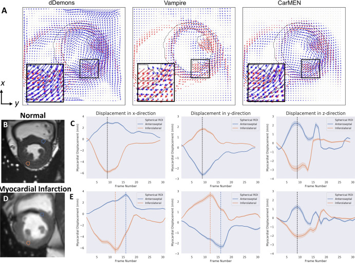
A, Representative images of synthetic motion estimate in-plane components. Predicted estimates (blue) are compared with ground truth (red). B-E, Example of assessment of left ventricle dyssynchrony with cardiac motion estimation network (CarMEN) in, B, C, a healthy individual and, D,E, a patient with myocardial infarction. B, Cardiac MR image in short-axis orientation. Sample spherical volumes are placed at anteroseptal (AS) and inferolateral (IL) walls near base. C, Corresponding displacement graphs show absence of left ventricle dyssynchrony as both AS and IL walls reach peak displacement simultaneously (black dotted lines). D, E, In a patient with myocardial infarction, IL wall reaches peak displacement four frames earlier (orange dotted lines) relative to AS wall (blue dotted lines) corresponding to approximately 120 msec for frame resolution of 30 msec. This difference may indicate left ventricle dyssynchrony related to infarcted tissue in septal wall. dDemons = Diffeomorphic Demons, ROI = region of interest.

