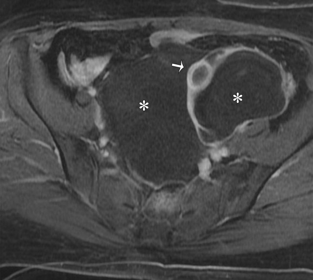
Figure 10b.
Peritoneal inclusion cyst. (a) Coronal T2-weighted image shows a large hyperintense lesion (*) surrounding the ovary (arrow). The ovary is entrapped by peritoneal adhesions and suspended centrally, producing a characteristic “spider in a web” appearance. (b) Contrast-enhanced T1-weighted image shows smooth mild enhancement, which represents adhesions. Solid enhancing nodules are absent. Note the normal enhancement of the ovary (arrow). * = cyst.

