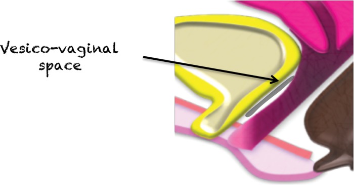
Figure 21a.
(a) Midsagittal plane through the female pelvis shows the vesicovaginal fascia. (b, c) Sagittal (b) and axial (c) T2-weighted images show an intermediate-SI lesion (arrow) in the vesicovaginal fascia. Note the mass effect on the urethra anteriorly and vagina posteriorly. At biopsy, the lesion represented vaginal sarcoma.

