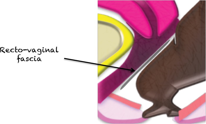
Figure 22a.
(a) Midsagittal plane through the female pelvis shows the rectovaginal fascia. (b, c) Sagittal (b) and axial (c) T2-weighted images show a large mass in the rectovaginal fascia (arrow in b), which communicates with the rectum (arrows in c). At biopsy, the mass was consistent with a GIST.

