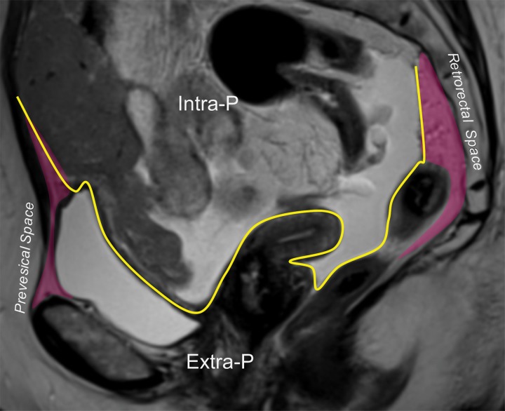
Figure 3b.
(a) Drawing of a midsagittal plane through the female pelvis shows the peritoneal reflection that divides the pelvis into the peritoneal and extraperitoneal compartments. (b) Sagittal T2-weighted MR image shows the anterior peritoneal reflection (yellow line). The presence of ascites makes it easy to identify the anterior peritoneal reflection. Extra-P = extraperitoneal compartment, Intra-P = intraperitoneal compartment.

