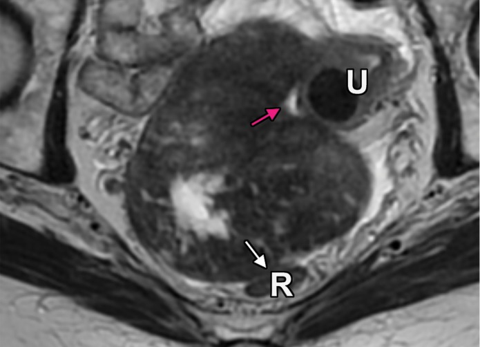
Figure 5b.
Masses arising from intraperitoneal organs or within the peritoneal cavity. R =rectum, U = uterus. (a) Axial T2-weighted image shows posterolateral displacement of the uterus (pink arrow) and lateral deviation of the external iliac vessels (white arrow) by a left ovarian mass. (b) Axial T2-weighted image shows an ovarian fibrothecoma in the rectouterine pouch displacing the uterus anteriorly (pink arrow) and rectum posteriorly (white arrow). (c, d) Imaging features of pelvic masses that arise from intraperitoneal organs. (c) Drawing shows posterior displacement of the uterus (pink arrow) by an intraperitoneal mass and lateral deviation of the external iliac vessels (white arrow). (d) Drawing shows a rectouterine mass displacing the uterus anteriorly (pink arrow) and rectum posteriorly (white arrow).

