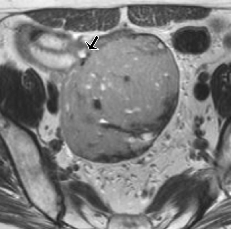
Figure 9b.
GIST. (a) Sagittal T2-weighted image shows a mass with intermediate SI (white arrow). Note that the mass is located above the anterior peritoneal reflection (black arrow). (b) Axial T2-weighted image shows that the mass arises from the ileum (arrow). (c) Axial contrast-enhanced fat-suppressed T1-weighted image shows poor enhancement of the mass (white arrow) and communication of the mass with the ileum (black arrow). Results of pathologic analysis were consistent with a GIST arising from the distal ileum.

