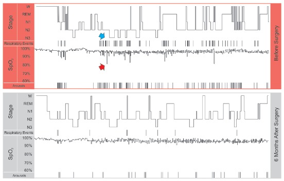
Figure 1. Segments of the polysomnographies of the patient presented in Figure 5, before and after orthognathic surgery. The hypnograms show the patient awake (W), and in the sleep stages of REM, 1 2 and 3. The respiratory events (apneas + hypopneas) are marked by vertical lines, as well as the microarousals (Arousals). Note that prior to surgery there is a certain association between microarousal and respiratory events. Also, blood oxygen saturation (SpO2) is associated with respiratory events; the red arrow points to one of the desaturation events that occurred after a respiratory event (blue arrow). The patient had important improvements in number of respiratory events, number of microarousals, blood oxygen saturation, and sleep quality with surgery.

